Definition
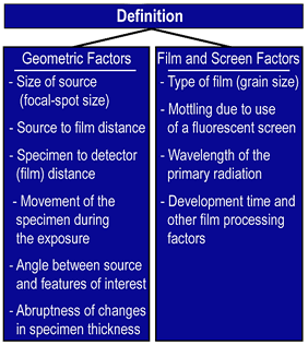
 As mentioned previously, radiographic definition is the abruptness of change from one density to another. Geometric factors of the equipment and the radiographic setup, and film and screen factors both have an effect on definition. Geometric factors include the size of the area of origin of the radiation, the source-to-detector (film) distance, the specimen-to-detector (film) distance, movement of the source, specimen or detector during exposure, the angle between the source and some feature and the abruptness of change in specimen thickness or density.
Geometric Factors
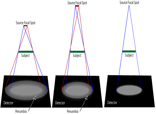
The effect of source size, source-to-film distance and the specimen-to-detector distance were covered in detail on the geometric unsharpness page. But briefly, to produce the highest level of definition, the focal-spot or source size should be as close to a point source as possible, the source-to-detector distance should be a great as practical, and the specimen-to-detector distance should be a small as practical. This is shown graphically in the images below.

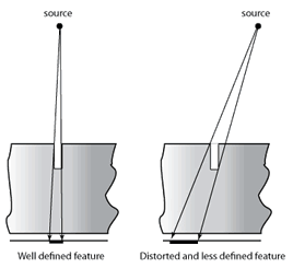
 The angle between the radiation and some features will also have an effect on definition. If the radiation is parallel to an edge or linear discontinuity, a sharp distinct boundary will be seen in the image. However, if the radiation is not parallel with the discontinuity, the feature will appear distorted, out of position and less defined in the image.
Abrupt changes in thickness and/or density will appear more defined in a radiograph than will areas of gradual change. For example, consider a circle. Its largest dimension will a cord that passes through its centerline. As the cord is moved away from the centerline, the thickness gradually decreases. It is sometimes difficult to locate the edge of a void due to this gradual change in thickness.
Lastly, any movement of the specimen, source or detector during the exposure will reduce definition. Similar to photography, any movement will result in blurring of the image. Vibration from nearby equipment may be an issue in some inspection situations.
Film and Screen Factors
The last set of factors concern the film and the use of fluorescent screens. A fine grain film is capable of producing an image with a higher level of definition than is a coarse grain film. Wavelength of the radiation will influence apparent graininess. As the wavelength shortens and penetration increases, the apparent graininess of the film will increase. Also, increased development of the film will increase the apparent graininess of the radiograph.
The use of fluorescent screens also results in lower definition. This occurs for a couple of different reasons. The reason that fluorescent screens are sometimes used is because incident radiation causes them to give off light that helps to expose the film. However, the light they produce spreads in all directions, exposing the film in adjacent areas, as well as in the areas which are in direct contact with the incident radiation. Fluorescent screens also produce screen mottle on radiographs. Screen mottle is associated with the statistical variation in the numbers of photons that interact with the screen from one area to the next.